
Longitudinal Iron Mapping in Alzheimer's Disease
Abnormally increased brain iron accumulation in deep gray matter is a common finding in Alzheimer’s disease (AD) and recent validation studies showed that brain iron can be measured precisely by quantitative MRI in vivo, thus, enabling reliable and precise longitudinal investigations.
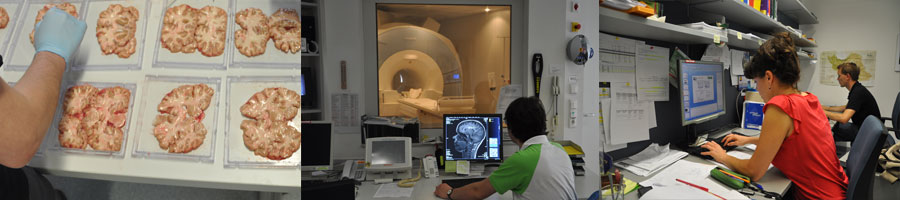
We conducted an explorative longitudinal study including 100 patients with AD and 100 age-matched control subjects, which were recruited for their 2 years follow-ups from our outpatient department and undergo extensive cognitive testing and quantitative 3 Tesla MRI. Regional differences of iron in deep gray matter and the neocortex were evaluated and related to the patients´ cognitive status. Follow-up MRI and clinical data with multivariate regression analysis served to investigate the dynamics of AD-related changes of susceptibility, and their relation to cognitive functioning.
We found higher iron concentration in the deep gray matter and neocortical regions in Alzheimer disease compared to healthy controls. Change in iron levels over time in the temporal lobe were associated with cognitive decline in individuals with AD.
Paper: https://doi.org/10.1148/radiol.2020192541
Press release " Brain Iron Accumulation Linked to Cognitive Decline in Alzheimer’s Patients" by RSNA Media Relations.
This research was funded by the Austrian Science Fund FWF KLIF 523.
References
** Damulina A, Pirpamer L, Soellradl M, Sackl M, Tinauer C, Hofer E, Enzinger C, Gesierich B, Duering M, Ropele S, Schmidt R, Langkammer C.
Cross-sectional and Longitudinal Assessment of Brain Iron in Alzheimer Disease using 3T MRI.
Radiology. 2020 Sep;296(3):619-626.
PubMed
** Langkammer, C; Bredies, K; Poser, BA; Barth, M; Reishofer, G; Fan, AP; Bilgic, B; Fazekas, F; Mainero; C; Ropele, S
Fast Quantitative Susceptibility Mapping using 3D EPI and Total Generalized Variation.
Neuroimage. 2015 Feb 27.
PubMed
** Langkammer, C; Krebs, N; Goessler, W; Scheurer, E; Yen, K; Fazekas, F; Ropele, S
Susceptibility induced gray-white matter MRI contrast in the human brain.
Neuroimage. 2012; 59(2):1413-1419
** Langkammer, C; Schweser, F; Krebs, N; Deistung, A; Goessler, W; Scheurer, E; Sommer, K; Reishofer, G; Yen, K; Fazekas, F; Ropele, S; Reichenbach, JR
Quantitative susceptibility mapping (QSM) as a means to measure brain iron? A post mortem validation study.
Neuroimage. 2012; 62(3):1593-1599