
Quantitative susceptibility mapping
Quantitative susceptibility mapping (QSM) is a novel post-processing technique which allows the calculation of the bulk magnetic susceptibility distribution of tissue in vivo from gradient echo magnetic resonance phase images. Susceptibility maps have been shown to demonstrate unprecedented anatomical contrast in both white and gray matter regions.
QSM reconstruction
QSM based on clinical data recently became feasible due to increased computational power and novel algorithms that enable both improved pre-processing of the input GRE data and reconstruction of susceptibility maps with reduced artifact level.
|
|
|
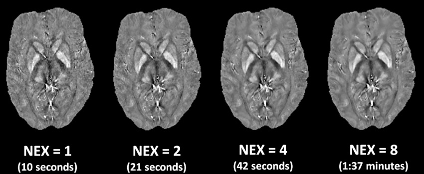
Figure: Reconstructed QSM images using total generalized variation (TGV). The QSM images have been acquired with a 3D EPI sequence (1mm isotropic resolution). Captions indicate the number of signal averages (NEX) and their respective acquisition times covering the entire brain. |
TGV QSM Algorithm
Source code for total generalized variation (TGV) based QSM can be downloaded at the QSM software page.
Biophysical effects underlying QSM
Recent experiments suggested relatively high correlation between tissue susceptibility assessed by QSM and the presumed regional
iron concentration. However, the bulk tissue magnetic susceptibility of white matter is also affected substantially by myelin, which is diamagnetic and, consequently, counteracts the effect of iron. In addition, substances such as blood and contributions from calcium and trace elements, have also been found to affect the magnetic susceptibility of tissue. Valid information on the true correlation of QSM measurements with tissue iron concentrations in different regions of the brain can, however, only be derived by direct comparison.
We investigate the relationship between brain
iron concentration,
myelin content,
white matter fiber orientation and magnetic susceptibility in unfixed (in situ)
postmortem brains scanned directly after death.
This research is funded by the Austrian Science Fund FWF.
Clinical applications of QSM
QSM provides information on the intrinsic biophysical tissue properties which is complementary to relaxation rate mapping. It may, therefore, be expected that the combination of relaxation rate mapping and QSM will provide further insight and will be instrumental for disentangling the main contributors yielding an increased accuracy of measurements of iron concentration and non-iron contributions in white matter.
The clinical potential of QSM is still under investigation but it is anticipated that susceptibility maps of patients will provide novel insights into disease induced tissue changes. First results in multiple sclerosis patients demonstrated that QSM is more sensitive to disease induced tissue changes than other quantitative MRI methods such as relaxation time mapping. Additionally, QSM allowed to assess tissue changes even in patients at a very early stage of MS.
References
** Langkammer, C; Schweser, F; Shmueli, K; Kames, C; Li, X; Guo, L; Milovic, C; Kim, J; Wei, H; Bredies, K; Buch, S; Guo, Y; Liu, Z; Meineke, J; Rauscher, A; Marques, JP; Bilgic, B
Quantitative susceptibility mapping: Report from the 2016 reconstruction challenge.
Magn Reson Med. 2018; 79(3):1661-1673
[Fulltext]
[Pubmed]
** Langkammer, C; Pirpamer, L; Seiler, S; Deistung, A; Schweser, F; Franthal, S; Homayoon, N; Katschnig-Winter, P; Koegl-Wallner, M; Pendl, T; Stoegerer, EM; Wenzel, K; Fazekas, F; Ropele, S; Reichenbach, JR; Schmidt, R; Schwingenschuh, P
Quantitative Susceptibility Mapping in Parkinson's Disease.
PLoS One. 2016; 11(9):e0162460-e0162460
[Fulltext]
[Pubmed]
** Birkl, C; Langkammer, C; Krenn, H; Goessler, W; Ernst, C; Haybaeck, J; Stollberger, R; Fazekas, F; Ropele, S
Iron mapping using the temperature dependency of the magnetic susceptibility.
Magn Reson Med. 2015; 73(3): 1282-1288.
[Fulltext]
[Pubmed]
** Langkammer, C; Bredies, K; Poser, BA; Barth, M; Reishofer, G; Fan, AP; Bilgic, B; Fazekas, F; Mainero, C; Ropele, S
Fast quantitative susceptibility mapping using 3D EPI and total generalized variation.
Neuroimage. 2015; 111(4): 622-630.
[Fulltext]
[Pubmed]
** Langkammer, C; Liu, T; Khalil, M; Enzinger, C; Jehna, M; Fuchs, S; Fazekas, F; Wang, Y; Ropele, S
Quantitative susceptibility mapping in multiple sclerosis.
Radiology. 2013; 267(2):551-559
[Fulltext]
[Pubmed]
** Damulina, A; Pirpamer, L; Soellradl, M; Sackl, M; Tinauer, C; Hofer, E; Enzinger, C; Gesierich, B; Duering, M; Ropele, S; Schmidt, R; Langkammer, C
Cross-sectional and Longitudinal Assessment of Brain Iron Level in Alzheimer Disease Using 3-T MRI.
Radiology. 2020; 296(3):619-626
[Fulltext]
[Pubmed]
** Langkammer, C; Krebs, N; Goessler, W; Scheurer, E; Yen, K; Fazekas, F; Ropele, S
Susceptibility induced gray-white matter MRI contrast in the human brain.
Neuroimage. 2012; 59(2): 1413-1419.
[Fulltext]
[Pubmed]
** Langkammer, C; Schweser, F; Krebs, N; Deistung, A; Goessler, W; Scheurer, E; Sommer, K; Reishofer, G; Yen, K; Fazekas, F; Ropele, S; Reichenbach, JR
Quantitative susceptibility mapping (QSM) as a means to measure brain iron? A post mortem validation study.
Neuroimage. 2012; 62(3):1593-1599
[Fulltext]
[Pubmed]