Validation of Diffusion MRI
Diffusion tensor imaging (DTI) and tractography reveal structural details of neuronal tissue in-vivo, which formerly were only accessable by histological studies following autopsy.
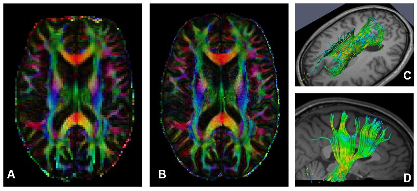
|
|
| Figure: Due to sequence design, the resolution of conventional DTI is limited to approx. 2 x 2 mm² in-plane (A). With readout-segmented sequences this restriction can be overcome and better resolutions are possible in-vivo (B, 1 x 1 mm²). This high-res DTI data serves as input for fiber tracking algorithms (C,D), which thus enable finer tractographical investigations in smaller anatomical structures. |
What are we measuring with DTI? - A postmortem validation
DTI and tractography are valuable tools which are used to gather further insight into brain microstructure and disease related tissue changes. However, relatively little is know about the biophysical mechanisms which are responsible for the generation of diffusion-weighted MRI and, thus, derived measures such as fractional anisotropy (FA) and mean diffusivity (MD).
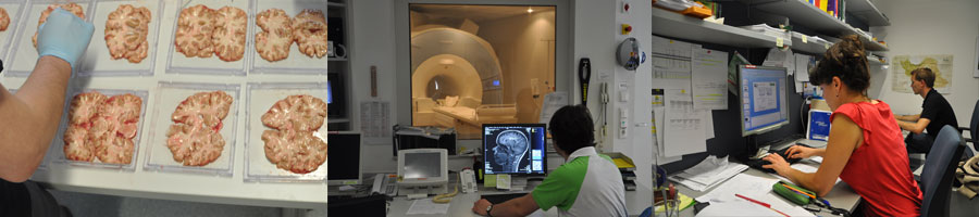
While investigations in formalin-fixed tissue samples are hampering further MRI measurements, we investigate postmortem brains in-situ (scanning the entire corpses). In this setup the tissue remains in condition comparable to in-vivo, but at a lower temperature. After correcting for effect of temperature, the diffusion processes can be modeled and compared to the amounts of specific lipids and proteins as well as to histological staining.
|
|
| Figure: (A) Feasibility of postmortem fiber tracking/tractography. (B) Dissection of the corpus callosum. In this example, tissue specimens are taken from a coronal brain slice and are subsequently analysed with lipidomic and proteomic measurement techniques. Combined with histological staining this serves to explain the generation of diffusion-derived MRI contrasts. |

References
** Langkammer, C; Krebs, N; Goessler, W; Scheurer, E; Fazekas, F; Ropele, S
Iron and myelin induced contrast variations in the corpus callosum.
21st Annual Meeting and Exhibition of the ISMRM; APR 20-26, 2013
** Langkammer, C; Enzinger, C; Quasthoff, S; Grafenauer, P; Soellinger, M; Fazekas, F; Ropele, S
Mapping of iron deposition in conjunction with assessment of nerve fiber tract integrity in amyotrophic lateral sclerosis.
J Magn Reson Imaging. 2010; 31(6): 1339-1345
** Langkammer, C; Krebs, N; Scheurer, E; Steiner, M; Fazekas, F; Yen, K; Stollberger, R; Ropele, S
Diffusion tensor imaging and tractography in post mortem brain tissue.
16th Annual Meeting of the Organization for Human Brain Mapping; JUN 6-10, 2010