
Neuromuscular Imaging
Outline
Neuromuscular disorders encompass a wide range of conditions affecting muscles, peripheral nerves, and the neuromuscular junction. These include, but are not limited to, muscular dystrophies, inflammatory myopathies, motor neuron diseases, and congenital myopathies. Accurate assessment of disease progression is essential for understanding pathophysiology and developing effective therapies.
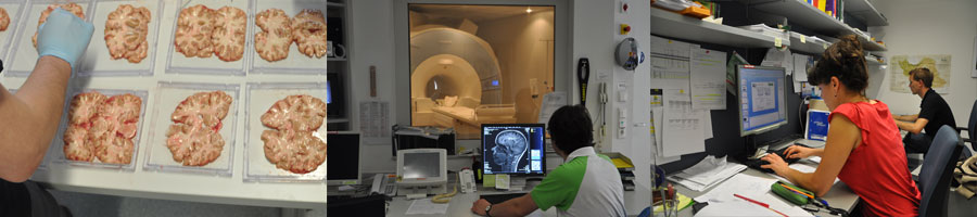
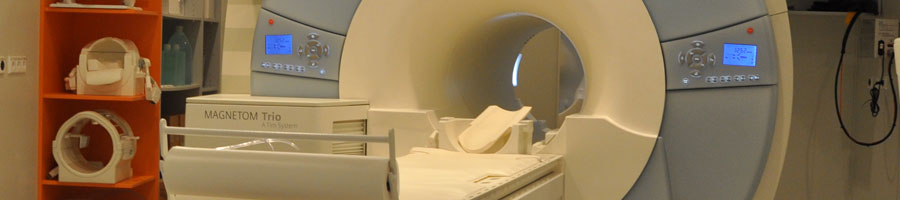
Our research unit focuses on advanced MRI techniques to noninvasively evaluate structural, metabolic, and functional changes in patients with neuromuscular disorders. By detecting early alterations in muscle tissue and quantifying progressive fat infiltration, we aim to establish sensitive imaging biomarkers that can guide therapeutic studies and clinical decision-making.
In addition to MRI, we integrate clinical evaluations, functional testing, and biochemical biomarkers from body fluids, providing a comprehensive overview of disease status. Our work is conducted in close collaboration with neurologists, physiotherapists, and other research units at the Medical University of Graz, fostering a multidisciplinary approach that bridges imaging, clinical care, and translational research.
Through this integrated strategy, we strive to improve diagnosis, monitor disease progression, and ultimately support the development of more effective treatments for a broad spectrum of neuromuscular disorders.
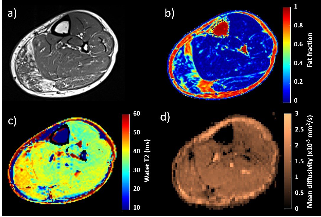
This figure shows MRI images of the lower leg from a patient with muscular dystrophy. Different MRI techniques allow us to visualize and analyze disease-related changes in muscle tissue:
- (a) Fat Replacement: Healthy muscle tissue is progressively replaced by fat, which can be detected and measured using MRI.
- (b) Fat Quantification: By analyzing MRI data, we can calculate the fat fraction in the muscle, providing an objective measure of disease progression.
- (c) Inflammation Detection: Some MRI techniques can reveal inflammation within the muscle, which may be an indicator of ongoing disease activity.
- (d) Structural Changes: MRI can also highlight alterations in muscle structure, helping to assess the overall integrity of the tissue.
By utilizing different MRI contrasts, we can obtain a comprehensive picture of disease progression, aiding in diagnosis, monitoring, and the evaluation of potential treatments.
Join Our Neuromuscular Imaging Research
Explore cutting-edge MRI studiesin neuromuscular disorders, from muscular dystrophies to motor neuron diseases. We combine advanced imaging with clinical and biochemical assessments to track disease progression and develop sensitive biomarkers.
With our upcoming CimaX scanner, we will pioneer multinuclear MRI (sodium & phosphorus) and work on standardizing muscle MRI protocols. Stay tuned for updates and opportunities to get involved!
Contact: Teresa Gerhalter
We welcome inquiries from students, PhD candidates, PostDocs, and collaborators interested in joining our research.